
A variety of cell studies including organelle detection and studies on function and proliferation of cells can be done using BioActs’ bright fluorescent probes and fluorescent antibodies. Since organelles have their own unique features, organelle-specific image tracking and analysis are very important research tools in cell biology. BioActs’ fluorescent probes for cell analysis purposes can be applied for live cell imaging, as well as fluorescent microscopy and flow cytometry.
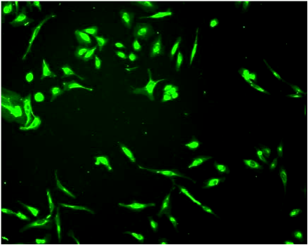
CytoFlamma Cell Membrane
CytoFlamma® Fluors are hydrophobic dyes that can be used for cell membrane labeling by directly penetrating into cell membranes. Because of its low cytotoxicity and ability to penetrate into cell membranes, it also can be used in live cells.
| Product Name | Product Code | Excitation/Emission (nm) |
|---|---|---|
| CytoFlamma 496 Cell-membrane (Live) | BCT-RCS5001 | 496 / 516nm |
| CytoFlamma 552 Cell-membrane (Live) | BCT-RCS1001 | 550 / 565nm |
| CytoFlamma 648 Cell-membrane (Live) | BCT-RCS1611 | 648 / 663nm |
| CytoFlamma 675 Cell-membrane (Live) | BCT-RCS3001 | 675 / 698nm |
| CytoFlamma 749 Cell-membrane (Live) | BCT-RCS1611 | 774 / 806nm |
| CytoFlamma ICG Cell-membrane (Live) | BCT-RCS1811 | 749 / 774nm |
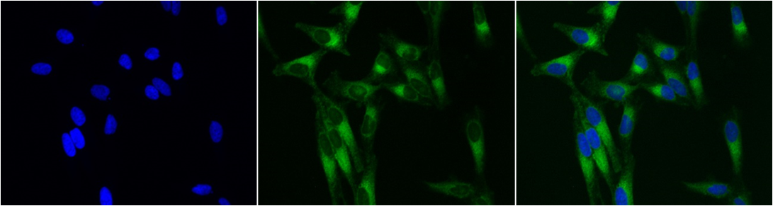
MitoFlamma Green
MitoFlamma™ Green is a green fluorescence probe specifically staining mitochondria of cells. By marking mitochondria of live cells, it can be used to trace cell mutations from drug or external stimuli. It can also be used for multi-imaging staining of fixed mitochondria. This probe can be applied for fluorescence microscopy or flow cytometry and analysis based on microplates.
MitoFlamma Green (Live) (BCT-RMS110), 508/580nm
ApoFlamma Dyes
ApoFlamma® dyes are developed for fluorescence imaging of apoptosis. There are two types of products groups, I) ApoFlamma® PS and II) ApoFlamma® H. The ApoFlamma® PS panel targets specifically to phosphatidylserine (PS) exposed on the cell-surface of dead cells and ApoFlamma® H panel of dyes specifically targets histone 1 exposed on the surface of apoptotic cells. A broad coverage of the whole spectra from UV to NIR allows a broad application range, including in vitro and in vivo studies.
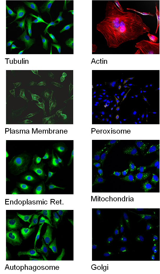
Organelle and Cell Target-specific Imaging
BioActs supplies cell target-specific primary antibodies and other probes. In order to get clear-cut images by using primary
 antibodies specific for cell organelle targets such as Tubulin, Mitochondria, Autophagosome, Golgi, ER or Peroxisomes, it is important to apply secondary
antibodies with excellent fluorescent dyes. BioActs’ fluorescent secondary antibodies provide best results in fluorescence microscopy, confocal laser scanning microscope (CLSM), flow cytometry or western blot. BioActs’ HRP secondary antibodies show ultimate performance in all areas of chemiluminescent detection systems as well.
| Product Description | Product Code | Target |
|---|---|---|
| Alpha Tubulin Antibody | BCT-RPA5414 | Tubulin |
| Flamma Phalloidin | 5 Products | Actin |
| CytoFlamma Dyes | 6 Products | Plasma Membrane |
| COX4 Antibody | BCT-RPA4111 | Mitochondria |
| Golgin-97 Antibody | BCT-RPA5212 | Golgi |
| LC3B Antibody | BCT-RPA4313 | Autophagosome |
| PMP70 Antibody | BCT-RPA4515 | Peroxisome |
| PDI Antibody | BCT-RPA5616 | Endoplasmic Reticulum |
Excellent Labeled Secondary Antibodies
- Goat anti-mouse IgG Secondary Antibodies
- Goat anti-rabbit IgG Secondary Antibodies
- Goat anti-rat IgG Secondary Antibodies