ACE2 Antibody

ProSci
Product Code: PSI-3229
Product Group: Primary Antibodies
Supplier: ProSci
CodeSizePrice
PSI-3229-0.02mg0.02mg£150.00
Quantity:
PSI-3229-0.1mg0.1mg£449.00
Quantity:
Prices exclude any Taxes / VAT

Overview

Host Type: Rabbit
Antibody Isotype: IgG
Antibody Clonality: Polyclonal
Regulatory Status: RUO
Applications:
  • Enzyme-Linked Immunosorbent Assay (ELISA)
  • Immunofluorescence (IF)
  • Immunohistochemistry (IHC)
  • Western Blot (WB)

Images

1 / 10
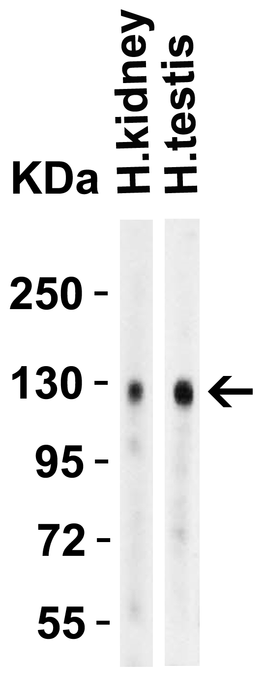
<strong>Figure 1 Western Blot Validation in Human Tissues</strong><br>
Loading: 15 ug of lysates per lane.
Antibodies: ACE2, 3229 (4 μg/mL),  1h incubation at RT  in 5% NFDM/TBST.
Secondary: Goat anti-rabbit IgG HRP conjugate at 1:10000 dilution.
2 / 10
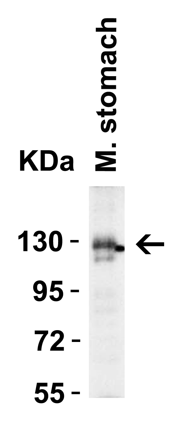
<strong>Figure 2 Western Blot Validation in Mouse Stomach Tissue</strong><br>
Loading: 15 μg of lysates per lane.
Antibodies: ACE2, 3229 (4 μg/mL),  1h incubation at RT  in 5% NFDM/TBST.
Secondary: Goat anti-rabbit IgG HRP conjugate at 1:10000 dilution.
3 / 10
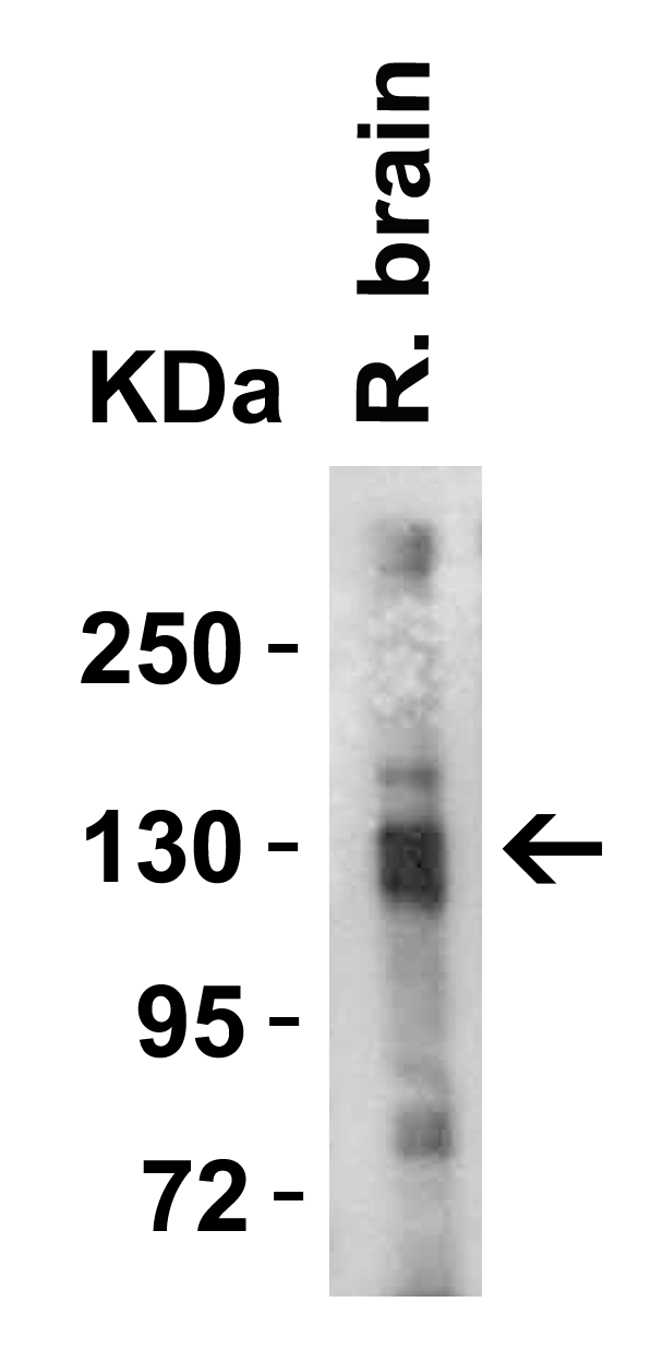
<strong>Figure 3 Western Blot Validation in Rat Brain Tissue</strong><br>
Loading: 15 μg of lysates per lane.
Antibodies: ACE2, 3229 (4 μg/mL),  1h incubation at RT  in 5% NFDM/TBST.
Secondary: Goat anti-rabbit IgG HRP conjugate at 1:10000 dilution.
4 / 10
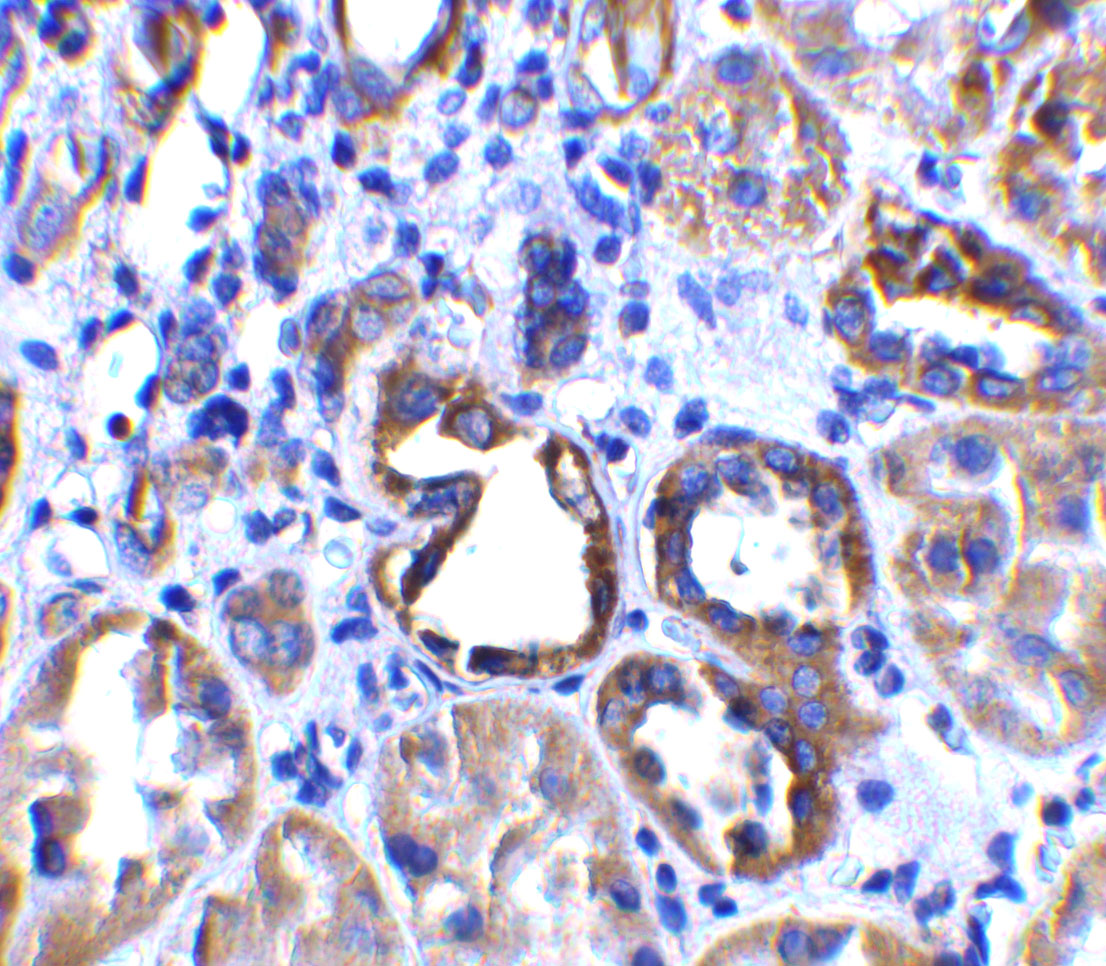
<strong>Figure 4 Immunohistochemistry Validation of ACE2 in Human Kidney Tissue </strong><br> 
Immunohistochemical analysis of paraffin-embedded human kidney tissue using anti-ACE2 antibody (3229) at 2 μg/ml. Tissue was fixed with formaldehyde and blocked with 10% serum for 1 h at RT; antigen retrieval was by heat mediation with a citrate buffer (pH6). Samples were incubated with primary antibody overnight at 4 ˚C. A goat anti-rabbit IgG H&L (HRP) at 1/250 was used as secondary. Counter stained with Hematoxylin.
5 / 10
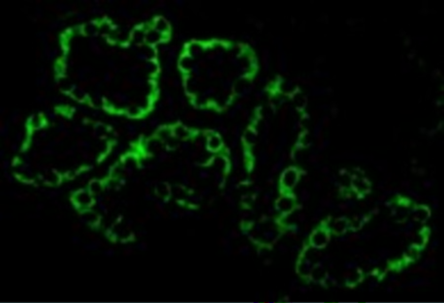
<strong>Figure 5 Immunofluorescence Validation of ACE2 in Human Kidney Cells</strong><br>
Immunofluorescent analysis of 4% paraformaldehyde-fixed human kidney cells labeling ACE2 with 3229 at 20 μg/mL, followed by goat anti-rabbit IgG secondary antibody at 1/500 dilution (green).
6 / 10
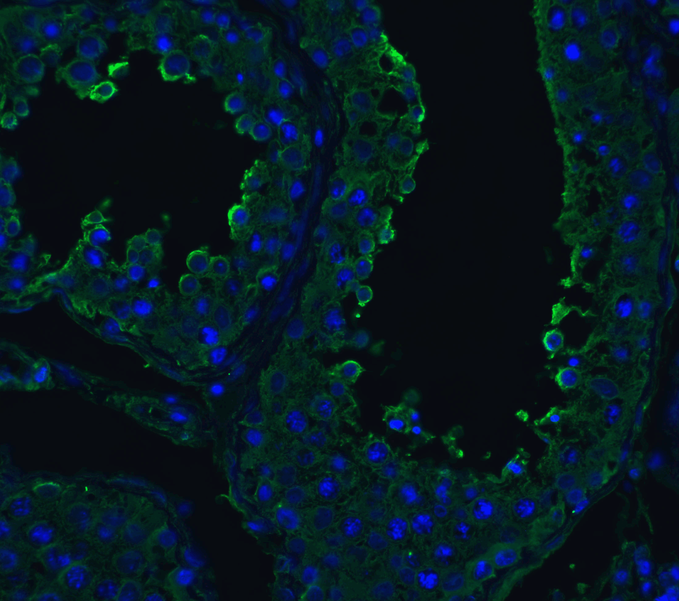
<strong>Figure 6 Immunofluorescence Validation of ACE2 in Human Testis Tissue</strong><br>
Immunofluorescent analysis of 4% paraformaldehyde-fixed human testis tissue labeling ACE-2 with 3229 at 20 μg/mL, followed by goat anti-rabbit IgG secondary antibody at 1/500 dilution (green) and DAPI staining (blue).
7 / 10
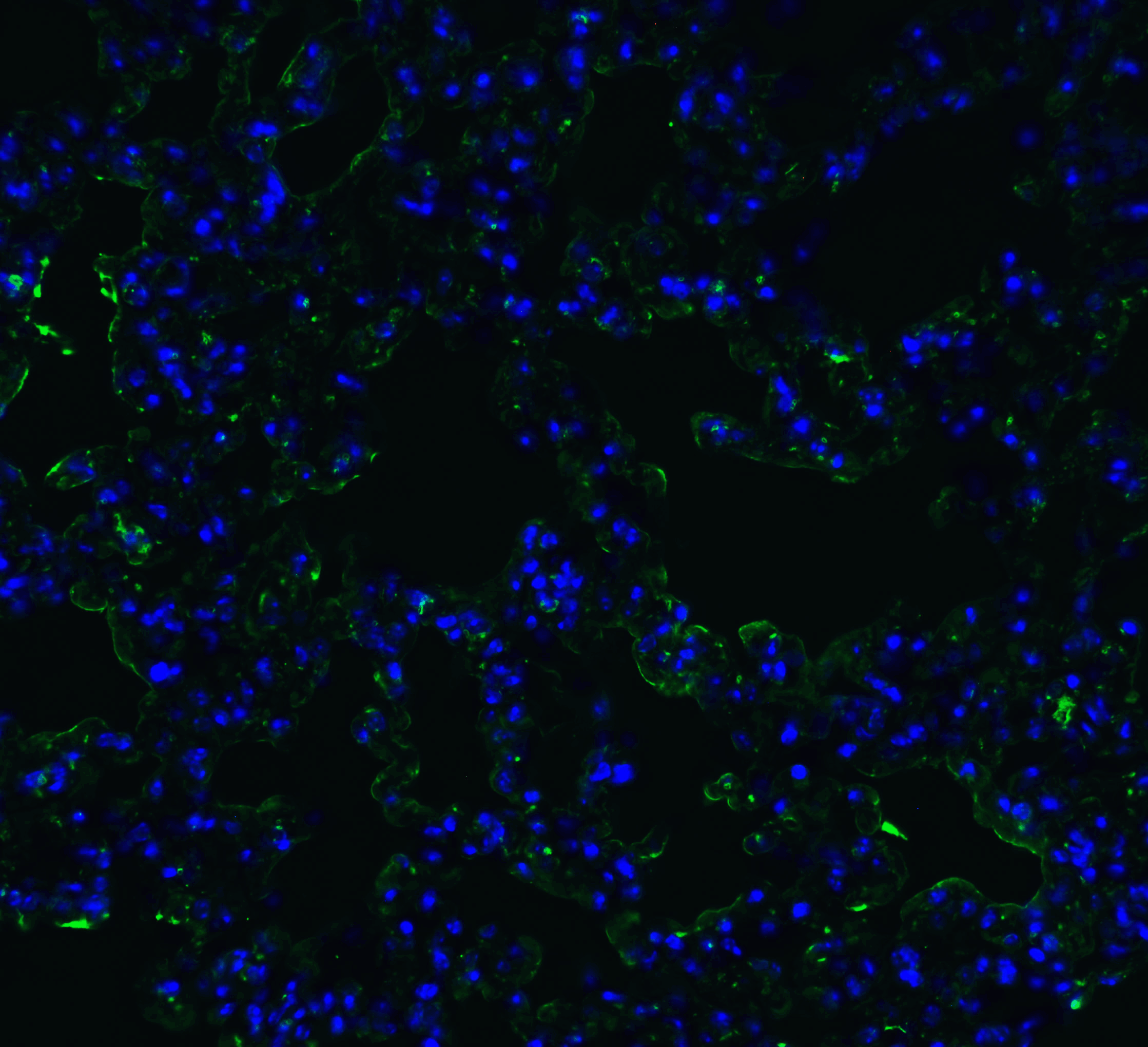
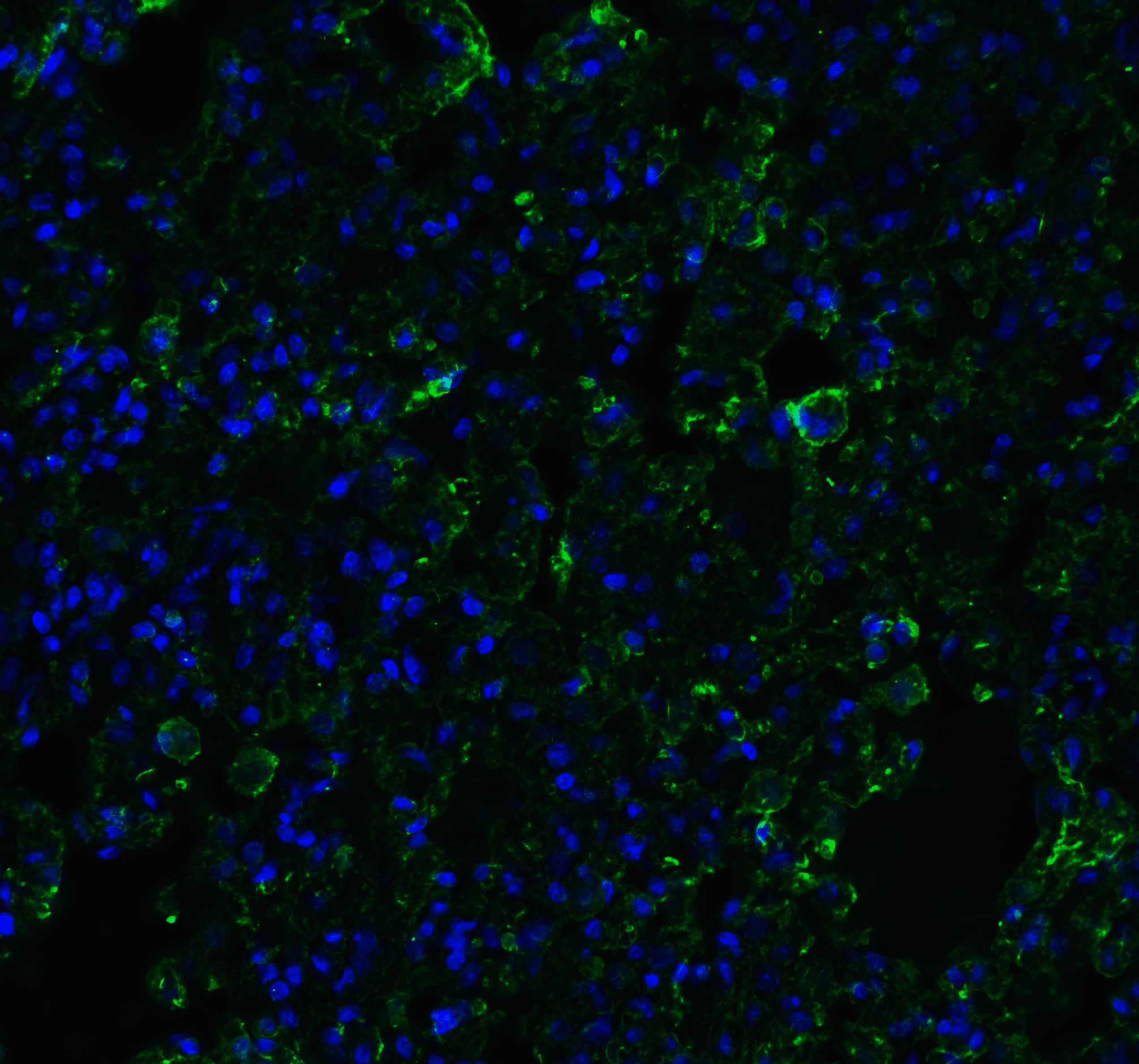
<strong>Figure 7 Immunofluorescence Validation of ACE2 in Human Lung Tissue</strong><br>
Immunofluorescent analysis of 4% paraformaldehyde-fixed human lung tissue labeling ACE-2 with 3229 at 20 μg/mL, followed by goat anti-rabbit IgG secondary antibody at 1/500 dilution (green) and DAPI staining (blue).
8 / 10
<strong>Figure 8 Immunofluorescence Validation of ACE2 in Mouse Lung Tissue</strong><br>
Immunofluorescent analysis of 4% paraformaldehyde-fixed mouse lung tissue labeling ACE-2 with 3229 at 20 μg/mL, followed by goat anti-rabbit IgG secondary antibody at 1/500 dilution (green) and DAPI staining (blue).
9 / 10
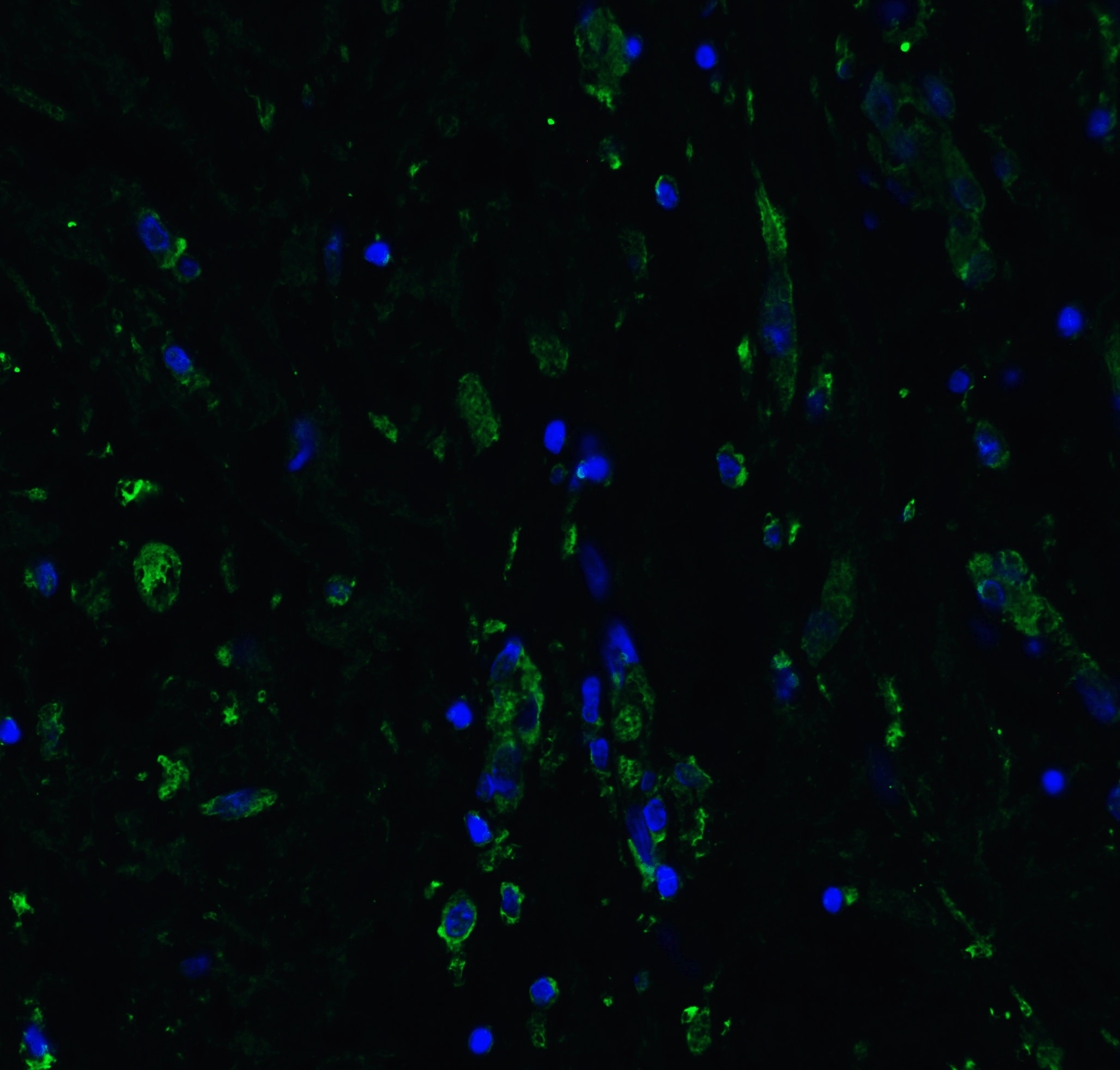
<strong>Figure 9 Immunofluorescence Validation of ACE2 in Rat Lung Tissue</strong><br>
Immunofluorescent analysis of 4% paraformaldehyde-fixed rat lung tissue labeling ACE-2 with 3229 at 20 μg/mL, followed by goat anti-rabbit IgG secondary antibody at 1/500 dilution (green) and DAPI staining (blue).
10 / 10
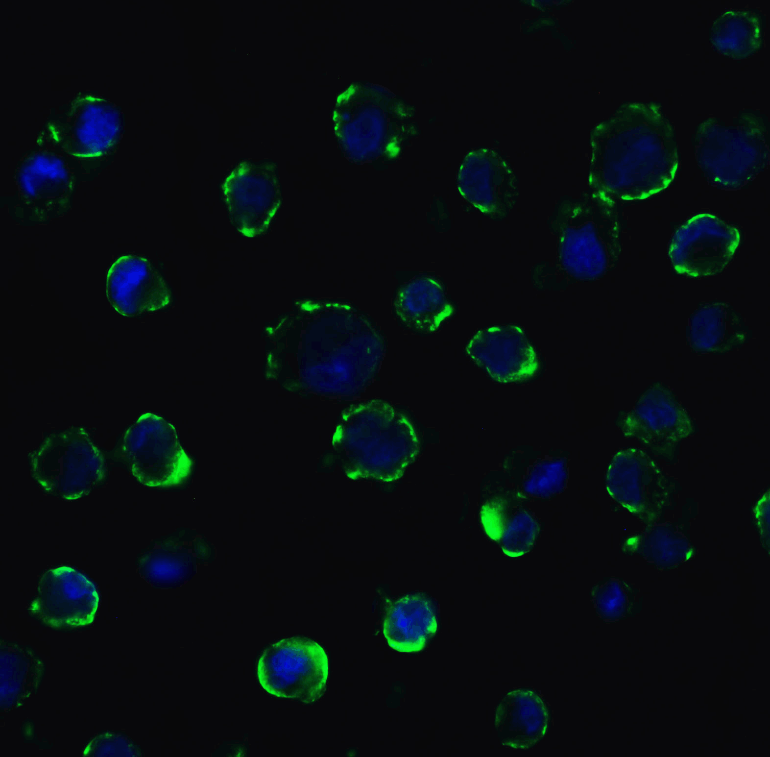
<strong>Figure 10 Immunofluorescence Validation of ACE2 In Caco2 Cells </strong><br> 
Immunofluorescent analysis of 4% paraformaldehyde-fixed Caco2 cells labeling ACE2 with 3229 at 5 μg/mL, followed by goat anti-rabbit IgG secondary antibody at 1/500 dilution (green) and DAPI staining (blue).  Image showing membrane staining on Caco2 cells.

<strong>Figure 1 Western Blot Validation in Human Tissues</strong><br>
Loading: 15 ug of lysates per lane.
Antibodies: ACE2, 3229 (4 μg/mL),  1h incubation at RT  in 5% NFDM/TBST.
Secondary: Goat anti-rabbit IgG HRP conjugate at 1:10000 dilution.
<strong>Figure 2 Western Blot Validation in Mouse Stomach Tissue</strong><br>
Loading: 15 μg of lysates per lane.
Antibodies: ACE2, 3229 (4 μg/mL),  1h incubation at RT  in 5% NFDM/TBST.
Secondary: Goat anti-rabbit IgG HRP conjugate at 1:10000 dilution.
<strong>Figure 3 Western Blot Validation in Rat Brain Tissue</strong><br>
Loading: 15 μg of lysates per lane.
Antibodies: ACE2, 3229 (4 μg/mL),  1h incubation at RT  in 5% NFDM/TBST.
Secondary: Goat anti-rabbit IgG HRP conjugate at 1:10000 dilution.
<strong>Figure 4 Immunohistochemistry Validation of ACE2 in Human Kidney Tissue </strong><br> 
Immunohistochemical analysis of paraffin-embedded human kidney tissue using anti-ACE2 antibody (3229) at 2 μg/ml. Tissue was fixed with formaldehyde and blocked with 10% serum for 1 h at RT; antigen retrieval was by heat mediation with a citrate buffer (pH6). Samples were incubated with primary antibody overnight at 4 ˚C. A goat anti-rabbit IgG H&L (HRP) at 1/250 was used as secondary. Counter stained with Hematoxylin.
<strong>Figure 5 Immunofluorescence Validation of ACE2 in Human Kidney Cells</strong><br>
Immunofluorescent analysis of 4% paraformaldehyde-fixed human kidney cells labeling ACE2 with 3229 at 20 μg/mL, followed by goat anti-rabbit IgG secondary antibody at 1/500 dilution (green).
<strong>Figure 6 Immunofluorescence Validation of ACE2 in Human Testis Tissue</strong><br>
Immunofluorescent analysis of 4% paraformaldehyde-fixed human testis tissue labeling ACE-2 with 3229 at 20 μg/mL, followed by goat anti-rabbit IgG secondary antibody at 1/500 dilution (green) and DAPI staining (blue).
<strong>Figure 7 Immunofluorescence Validation of ACE2 in Human Lung Tissue</strong><br>
Immunofluorescent analysis of 4% paraformaldehyde-fixed human lung tissue labeling ACE-2 with 3229 at 20 μg/mL, followed by goat anti-rabbit IgG secondary antibody at 1/500 dilution (green) and DAPI staining (blue).
<strong>Figure 8 Immunofluorescence Validation of ACE2 in Mouse Lung Tissue</strong><br>
Immunofluorescent analysis of 4% paraformaldehyde-fixed mouse lung tissue labeling ACE-2 with 3229 at 20 μg/mL, followed by goat anti-rabbit IgG secondary antibody at 1/500 dilution (green) and DAPI staining (blue).
<strong>Figure 9 Immunofluorescence Validation of ACE2 in Rat Lung Tissue</strong><br>
Immunofluorescent analysis of 4% paraformaldehyde-fixed rat lung tissue labeling ACE-2 with 3229 at 20 μg/mL, followed by goat anti-rabbit IgG secondary antibody at 1/500 dilution (green) and DAPI staining (blue).
<strong>Figure 10 Immunofluorescence Validation of ACE2 In Caco2 Cells </strong><br> 
Immunofluorescent analysis of 4% paraformaldehyde-fixed Caco2 cells labeling ACE2 with 3229 at 5 μg/mL, followed by goat anti-rabbit IgG secondary antibody at 1/500 dilution (green) and DAPI staining (blue).  Image showing membrane staining on Caco2 cells.

Further Information

Additional Names:
ACE2 Antibody: ACEH, Angiotensin-converting enzyme 2, ACE-related carboxypeptidase, ACEH, SARS-CoV receptor, SARS-CoV-2 receptor
Application Note:
WB: 4 μg/mL; IHC: 2 μg/mL; IF: 20 μg/mL.

Antibody validated: Western Blot in human, mouse and rat samples; Immunohistochemistry in human, mouse and rat samples; Immunofluorescence in human, mouse, and rat samples. All other applications and species not yet tested.
Background:

ACE2 Antibody: Angiotensin-converting enzyme 2 (ACE2) plays a central role in vascular, renal, and myocardial physiology. In contrast to its homolog ACE, ACE2 expression is restricted to heart, kidney, and testis. Recently. ACE2 has also been shown to be a functional receptor of the SARS coronavirus. Homology modeling shows 2019-nCoV has a similar receptor-binding domain structure as SARS-CoV, which suggests COVID-19 (2019-nCoV) may use ACE2 as a receptor in humans for infection. The normal function of ACE2 is to convert the inactive vasoconstrictor angiotensin I (AngI) to Ang1-9 and the active form AngII to Ang1-7, unlike ACE, which converts AngI to AngII. While the role of these vasoactive peptides is not well understood, lack of ACE2 expression in ace2-/ace2- mice leads to severely reduced cardiac contractility, indicating its importance in regulating heart function.

Background References:
  • Donoghue et al. Circ. Res. 2000;87:1-9.
  • Tipnis et al. J Biol. Chem. 2000;275:33238-43.
  • Li et al. Nature 2003;426:450-4.
  • Lu et al. The Lancet 2020 (published online).
  • Crackower et al. Nature 2002;417:822-8.
Buffer:
ACE2 Antibody is supplied in PBS containing 0.02% sodium azide.
Concentration:
1 mg/mL
Conjugate:
Unconjugated
DISCLAIMER:
Optimal dilutions/concentrations should be determined by the end user. The information provided is a guideline for product use. This product is for research use only.
Homology:
Predicted species reactivity based on immunogen sequence: Bovine: (93%)
Immunogen:
ACE2 antibody was raised against a synthetic peptide corresponding to amino acids near the center of human ACE2.

The immunogen is located within amino acids 150 - 200 of ACE2.
ISOFORMS:
Human ACE2 has 2 isoforms, including isoform 1 (805aa, 93kD) and isoform 2 (555aa, 64kD). Mouse ACE2 also has 2 isoforms, including isoform 1 (805aa, 92kD) and isoform 2 (353aa, 40kD). Rat ACE2 has one isoform (805aa, 93kD). 3229 can detect human, mouse and rat.
NCBI Gene ID #:
59272
NCBI Official Name:
angiotensin I converting enzyme (peptidyl-dipeptidase A) 2
NCBI Official Symbol:
ACE2
NCBI Organism:
Homo sapiens
Physical State:
Liquid
PREDICTED MOLECULAR WEIGHT:
Predicted: 93kD

Observed: 130 kD (7 N-linked glycosylation)
Protein Accession #:
NP_068576
Protein GI Number:
11225609
Purification:
ACE2 Antibody is affinity chromatography purified via peptide column.
Research Area:
Infectious Disease
SPECIFICITY:
Anti-ACE2 has no cross response to ACE1.
Swissprot #:
Q9BYF1
User NOte:
Optimal dilutions for each application to be determined by the researcher.

Related Products

Product NameProduct CodeSupplier 
ACE2 (IN2) PeptidePSI-3229PProSci Summary Details