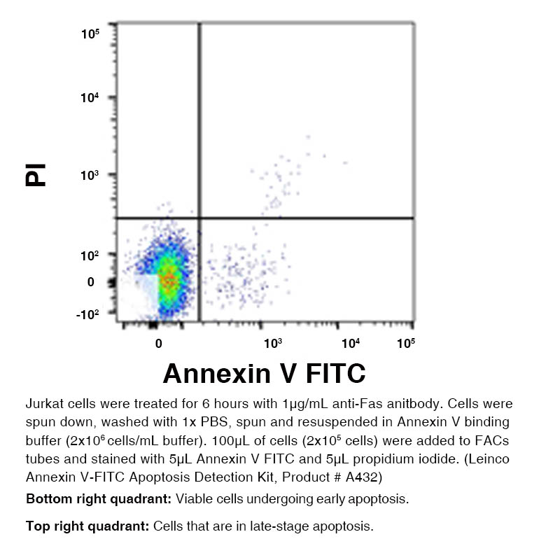
Annexin V - FITC Apoptosis Detection Kit

Leinco Technologies
Product Code: LEI-A432
Product Group: Cell Based Assays
CodeSizePrice
LEI-A432-25tests25 tests£151.00
Quantity:
LEI-A432-100tests100 tests£247.00
Quantity:
LEI-A432-300tests300 tests£408.00
Quantity:
Prices exclude any Taxes / VAT

Overview

Regulatory Status: RUO

Images

1 / 1

Further Information

Long Description:
Apoptosis, the physiological process of cell death, is a naturally occurring phenomenon. It is normally a tightly regulated process; however, if deregulated, tumor growth can result. Apoptotic processes can be measured in the laboratory easily and most effectively by using a FITC-labeled Annexin V protein to stain cells and analyze them via flow cytometry. It is a useful method to screen and analyze cells in a high throughput manner, resulting in a clearer understanding of the cellular response to various stimuli. When cells are undergoing cell death, phosphatidylserine (PS), a phospholipid which makes up a portion of the cell membrane, will translocate to the outside-facing side of the membrane. This translocation in the cell death mechanism exposes PS to the extracellular environment. Annexin V, a 35-36 kDa phospholipid-binding protein, has a high affinity for PS (KDa=7nmol/l). Once Annexin V is bound to the apoptotic cell it acts as a beacon for cells that have undergone apoptosis. Annexin V is often conjugated to FITC so the amount of signal can be measured and analyzed from labeled cells using a flow cytometer. The translocation of PS to the outside or exposed side of the membrane is an early event in the apoptotic process. Therefore, Annexin V staining is considered a marker for early stage apoptosis. Often times, Annexin V is paired with Propidium Iodide (PI) stain in order to analyze for late-apoptotic or dead cells. Early stage apoptotic cells will only take up the Annexin V stain but will remain PI negative, and late-stage apoptotic cells will be positive for both Annexin V and PI.
Target:
Annexin V

References

1. Piazza, GA. et al. (2016) Oncotarget. 7(5):5353-65.PubMed 2. Gassman, NR. et al. (2018) Environ Toxicol. 33(3):333-342.PubMed 3. Gelles, JD. and Chipuk, JE. (2016) Cell Death Dis. 7(12): e2493.