CycLex CaM-kinase II assay kit
| Code | Size | Price |
|---|
| MBL-CY-1173 | 96 Assays | £602.00 |
Quantity:
Prices exclude any Taxes / VAT
Overview
Regulatory Status: RUO
Shipping:
4°C
Storage:
4°C. Please refer to datasheet for additional information
Images
Documents
Further Information
Applications:
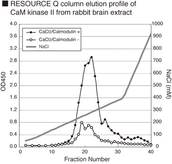
Other - 1) Monitoring the purification of CaM kinase II.
2) Screening inhibitors or activators of CaM kinase II
3) Detecting the effects of pharmacological agents on CaM kinase II activity.
Background:
Ca2+/CaM-dependent protein kinase II (CaM kinase II) is a ubiquitously expressed protein kinase that transduces elevated Ca2+ signals in cells to a number of target proteins ranging from ion channels to transcriptional activators. CaM kinase II has a unique holoenzyme structure and autoregulatory properties that allow it to give a prolonged response to transient Ca2+ signals and to sense cellular Ca2+ oscillations (1). In neurons CaM kinase II is highly expressed and localized with certain subcellular structures. Upon activation it can translocate to excitatory synapses where it regulates a number of proteins involved in synaptic transmission and its downstream signaling pathways. Changes in intracellular calcium can display variable responses ranging from highly localized, transient elevations within subcellular structures (e.g. a dendritic spine of a neuron) to Ca2+ waves that spread throughout the cell including the nucleus. The most ubiquitous calcium-sensing protein is Calmodulin (CaM), which contains four ?EF? hand motifs with high specificity for binding Ca2+. The Ca2+/CaM complex interacts with and modulates the functionality of a large number of proteins (2) including several Ser/Thr protein kinases. The CaM kinase II family is encoded by four genes (alpha, beta, gamma, and delta) that also exhibit alternative splicing. The gamma and delta isoforms are expressed in most tissues, whereas the alpha and beta isoforms are most prominent in neural tissues and comprise up to 2 % of the total protein in the hIPocampus of rodents and up to 1% of the total protein in the forebrain itself (3). The various CaM kinase II subunits are comprised of an N-terminal catalytic region, a central regulatory domain containing an autoinhibitory domain (AID) and Ca2+/CaM binding motif, a variable sequence, and the C-terminal subunit association domain (4). The holoenzyme is an oligomeric protein comprised of twelve 50?60-kDa subunits arranged as two stacked hexameric rings (5, 6). The C-terminal association domains form the central core of each ring with the N-terminal catalytic domains projecting outward. In the absence of bound Ca2+/CaM, the CaM kinase II is maintained in an inactive conformation because of an interaction of the AID with the catalytic domain of its own subunit. The Ca2+/CaM complex binds to a sequence that partially overlaps the AID, presumably causing a conformational change and thereby disrupting interaction of the AID with the catalytic domain and producing kinase activation. Interestingly, the sensitivity of CaM kinase II to activation by Ca2+/CaM depends on the subunit composition of the holoenzyme (7).
Description:
The CycLex® CaM kinase II Assay kit is primarily designed to measure the activities of purified
Ca2+/Calmodulin dependent protein kinase II (CaM kinase II) for the rapid and sensitive evaluation of
inhibitors or activators. The phospho-specific monoclonal antibody used in this assay kit has been
demonstrated to recognize the phospho-threonine residue in ?Syntide-2?, which is efficiently
phosphorylated by CaM kinase II. Additionally, column fractions of any cultured primary cell, cell line,
or tissue homogenate can be assayed for CaM kinase II activity with the CycLex® CaM kinase II Assay
kit if the appropriate dose of CaM kinase II specific inhibitor e.g. KN-62 is used.
Gene IDs:
Human: 816 Mouse: 12323
Kit Components:
Microplate, 10X Wash Buffer, Kinase Buffer, 50X CaCl2, 50X EGTA, 20X ATP, HRP conjugated Detection Antibody, Substrate Reagent, Stop Solution
Target:
CaMKII
References
1) Andy HUDMON1 and Howard SCHULMAN1 Biochem. J. 364, 593-611, 2002
(2) Van Eldik, L., and Watterson, M. Calmodulin and Signal Transduction, Academic Press, New York,
1998
(3) Erondu, N. E. and Kennedy, M. B. Regional distribution of type II Ca2+/calmodulin-dependent
protein kinase in rat brain. J. Neurosci. 5, 3270-3277, 1985
(4) Soderling, T. R. Biochim. Biophys. Acta 1297, 131?138, 1996
(5). Kolodziej, S. J., Hudmon, A., Waxham, M. N., and Stoops, J. K. J. Biol. Chem. 275, 14354?14359,
2000
(6). Kanaseki, T., Ikeuchi, Y., Sugiura, H., and Yamauchi, T. J. Cell Biol. 115, 1049-1060, 1991
(7) Brocke, L., Chiang, L. W., Wagner, P. D., and Schulman, H. J. Biol. Chem. 274, 22713?22722,
1999


