PDL-1 Antibody

ProSci
Product Code: PSI-4059
Product Group: Primary Antibodies
Supplier: ProSci
CodeSizePrice
PSI-4059-0.02mg0.02mg£150.00
Quantity:
PSI-4059-0.1mg0.1mg£449.00
Quantity:
Prices exclude any Taxes / VAT

Overview

Host Type: Rabbit
Antibody Isotype: IgG
Antibody Clonality: Polyclonal
Regulatory Status: RUO
Applications:
  • Enzyme-Linked Immunosorbent Assay (ELISA)
  • Immunofluorescence (IF)
  • Immunohistochemistry (IHC)
  • Western Blot (WB)

Images

1 / 15
<strong>Figure 1 Western Blot Validation of PD-L1 in HeLa Cells</strong><br>
Loading: 15 μg of lysates per lane.
Antibodies: 4059 (A: 1 μg/mL, B: 2 μg/mL), 1 h incubation at RT in 5% NFDM/TBST.
Secondary: Goat anti-rabbit IgG HRP conjugate at 1:10000 dilution.
2 / 15
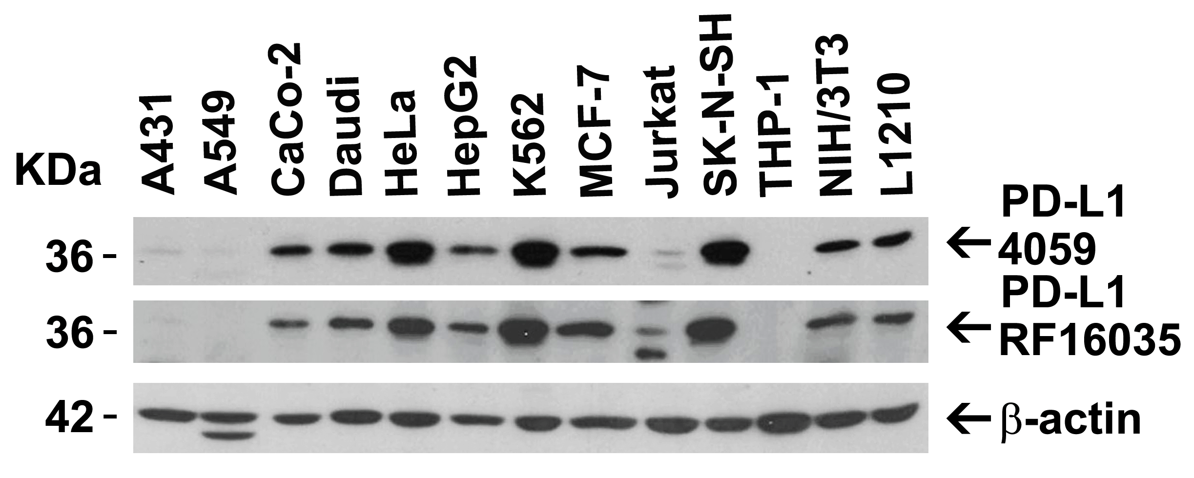
<strong>Figure 2 Independent Antibody Validation (IAV) via Protein Expression Profile in Human and Mouse cell lines</strong><br>
Loading: 15 μg of lysates per lane.
Antibodies: 4059 (2 μg/mL), RF16035 (2 μg/mL), and beta-actin (1 μg/mL), 1 h incubation at RT in 5% NFDM/TBST.
Secondary: Goat anti-rabbit and or anti-mouse IgG HRP conjugate at 1:10000 and 1:5000 dilution, respectively.
3 / 15
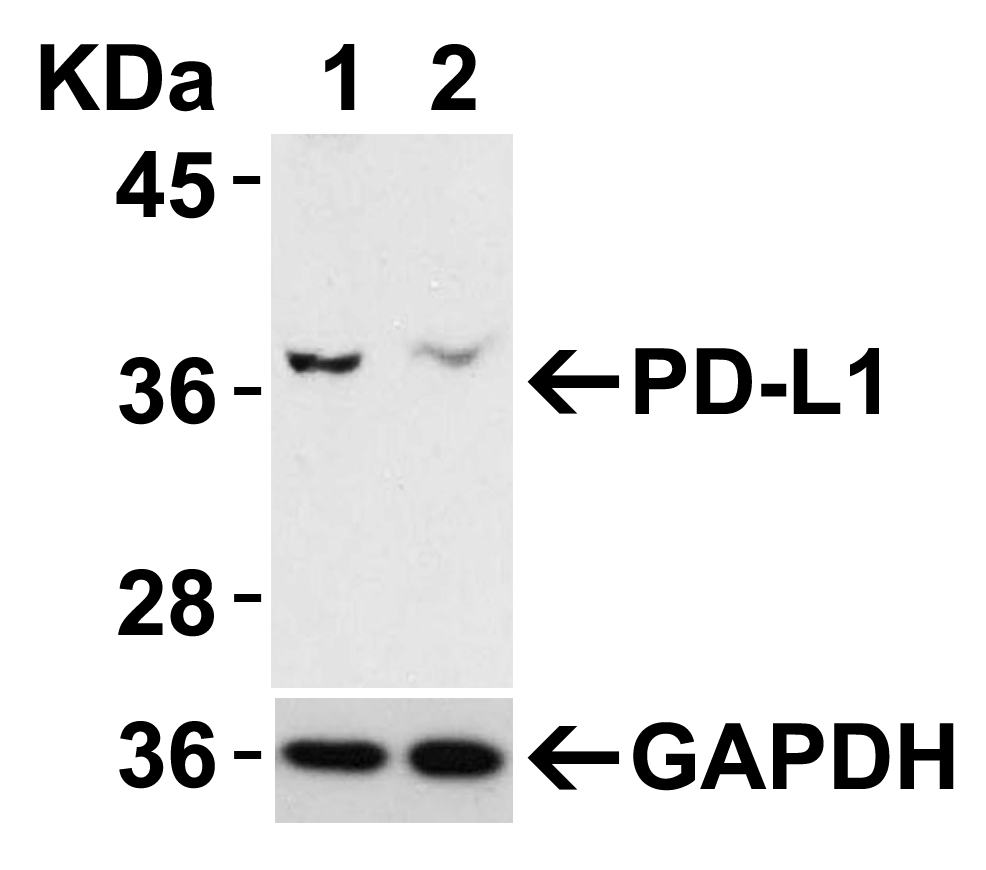
<strong>Figure 3 Validation with PD-L1 siRNA Knockdown in HeLa Cells</strong><br> 
HeLa cells were transfected with control siRNAs (lane 1) or PD-L1 siRNAs (lane 2) 
Loading: 10 μg of HeLa whole cell lysates per lane.
Antibodies: 4059 (2 μg/mL) and GAPDH (3783, 0.02 μg/mL), 1 h incubation at RT in 5% NFDM/TBST.
Secondary: Goat anti-mouse IgG HRP conjugate at 1:5000 dilution.
4 / 15
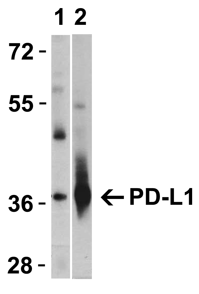
<strong>Figure 4 Validation with PD-L1 overexpression in 293 cells </strong><br> 
Loading: Lysates/proteins at 15 μg per lane.
Lane 1: non-transfected 293 cells
Lane 2: PD-L1 overexpressed 293 cells
Antibodies: 4059 (1 μg/mL). 1 h incubation at RT in 5% NFDM/TBST.
Secondary: Goat anti-rabbit IgG HRP conjugate at 1:10000 dilution.
5 / 15
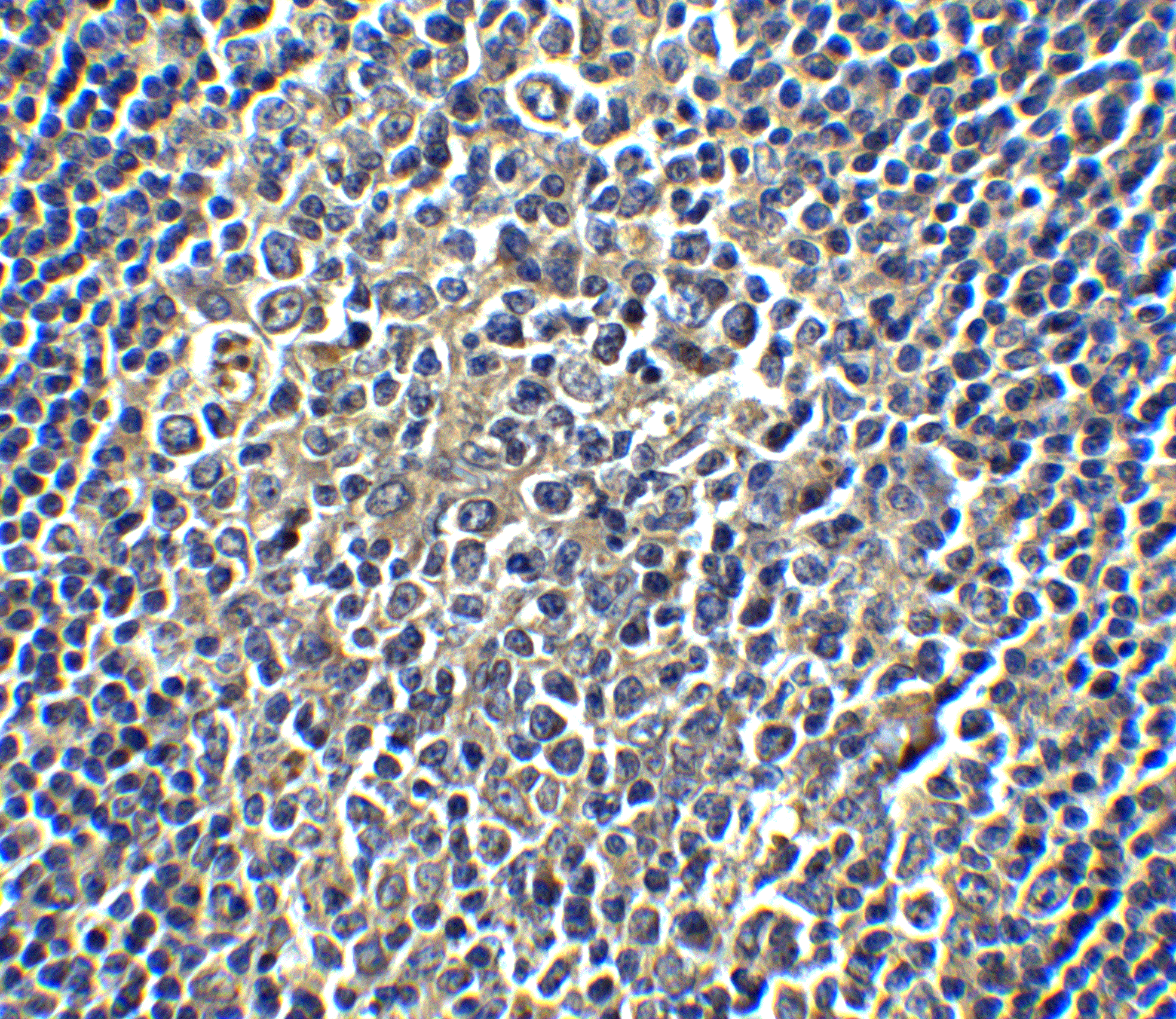
<strong>Figure 5 Immunohistochemistry Validation of PD-L1 in Human Tonsil Cells</strong><br> 
Immunohistochemical analysis of paraffin-embedded human tonsil tissue using anti-PD-L1 antibody (4059) at 5 μg/ml. Tissue was fixed with formaldehyde and blocked with 10% serum for 1 h at RT; antigen retrieval was by heat mediation with a citrate buffer (pH6). Samples were incubated with primary antibody overnight at 4˚C. A goat anti-rabbit IgG H&L (HRP) at 1/250 was used as secondary. Counter stained with Hematoxylin.
6 / 15
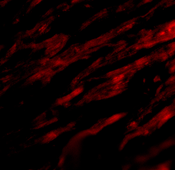
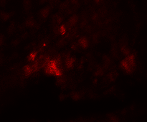
<strong>Figure 6 Immunofluorescence Validation of PD-L1 in Human Heart </strong><br>
Immunofluorescent analysis of 4% paraformaldehyde-fixed human heart tissue labeling PD-L1 with 4059 at 20 μg/mL, followed by goat anti-rabbit IgG secondary antibody at 1/500 dilution (red). Image showing both membrane and cytoplasmic staining on human heart tissue.
7 / 15
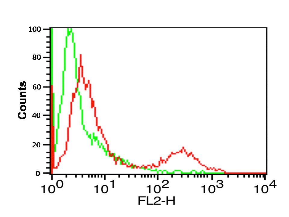
<strong>Figure 7 Flow Cytometry Validation of PD-L1 </strong><br>
Overlay histogram showing A-20 cells stained with 4059 (red line, 1μg/1x10<sup>6</sup> cells). 1 h incubation at 4˚C in 2% FBS/PBS. Followed by secondary antibody 488 goat anti-rabbit IgG (H+L) at 1/500 dilution for 1 h 4˚C. 
Isotype control antibody (Green line) was mouse IgG1 (1μg/1x10<sup>6</sup> cells) used under the same conditions. Acquisition of >10,000 events was performed.
8 / 15
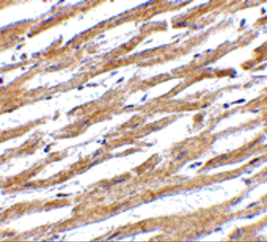
<strong>Figure 8 Immunohistochemistry Validation of PD-L1 in Rat Heart</strong><br>
Immunohistochemical analysis of paraffin-embedded rat heart tissue using anti-PD-L1 antibody (4059) at 5 μg/ml. Tissue was fixed with formaldehyde and blocked with 10% serum for 1 h at RT; antigen retrieval was by heat mediation with a citrate buffer (pH6). Samples were incubated with primary antibody overnight at 4˚C. A goat anti-rabbit IgG H&L (HRP) at 1/250 was used as secondary. Counter stained with Hematoxylin.
9 / 15
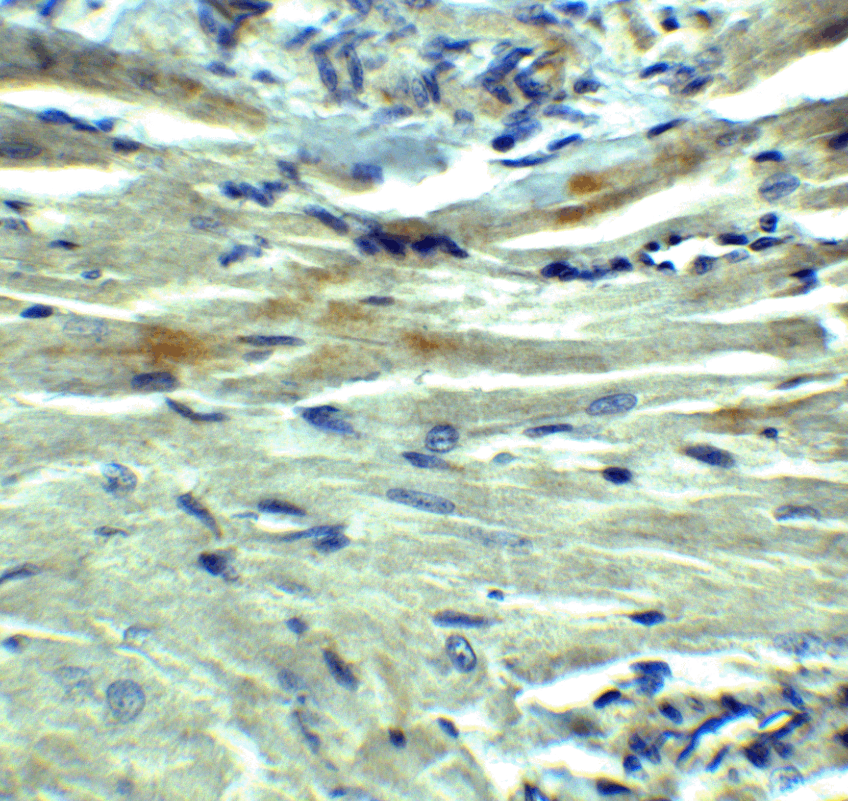
<strong>Figure 9 Immunohistochemistry Validation of PD-L1 in Human Heart</strong><br>
Immunohistochemical analysis of paraffin-embedded human heart tissue using anti-PD-L1 antibody (4059) at 2.5 ˚g/ml. Tissue was fixed with formaldehyde and blocked with 10% serum for 1 h at RT; antigen retrieval was by heat mediation with a citrate buffer (pH6). Samples were incubated with primary antibody overnight at 4˚C. A goat anti-rabbit IgG H&L (HRP) at 1/250 was used as secondary. Counter stained with Hematoxylin.
10 / 15
<strong>Figure 10 Immunofluorescence Validation of PD-L1 in Rat Heart</strong><br>
Immunofluorescence analysis of 4% paraformaldehyde-fixed rat heart tissue labeling PD-L1 with 4059 at 20 μg/ml, followed by goat anti-rabbit IgG secondary antibody at 1/250 dilution (red).
11 / 15
<strong>Figure 11 Immunofluorescence Validation of PD-L1 in tumors in Human Cells (Dhar et al., 2018) </strong><br>
(A) Several antibody brands were first tested with RBCs, WBCs, and HeLa cells: BioLegend, ProSci, and eBioscience. <strong>ProSci (4059) was chosen as it provided the highest staining intensity</strong><br>. (B, C) Using the optimal conditions of anti-PDL1 (ProSci Inc) at a concentration of 50μg/mL, following by goat anti-rabbit Alexa Fluor 647, PDL-1 staining was tested on several lung cancer cell lines: A549 (adenocarcinoma), H1703 (adenocarcinoma), H3255 (squamous) and WBCs as a control. (D) Once validated, patient samples were stained for PD-L1, CK, CD45, DAPI.
12 / 15
<strong>Figure 12 Immunohistochemistry Validation of PD-L1 in Human Tumors (Gadiot et al., 2011) </strong><br>
Immunohistochemical analysis of patient tumors labeling PD-L1 with anti-PD-L1 antibodies (4059). Several anti-PD-L1 antibodies were tested for staining, <strong> ?Only 1 antibody gave no background staining and was competitively blocked by the addition of PD-L1Fc protein (ProSci, #4059)?. <strong>
13 / 15
<strong>Figure 13 Immunohistochemistry Validation of PD-L1 in Human thyroid cancer (Angell et al., 2014) </strong><br>
Immunohistochemical analysis of PD-L1 expression in human thyroid cancer with anti-PD-L1 antibodeis (4059). Placenta was used a positive control.
14 / 15
<strong>Figure 14 Immunohistochemistry Validation of PD-L1 in Human Lung Cancer (Ilie et al., 2015) </strong><br>
Surgical specimens (left panel) and matched biopsy specimens (right panel). PD-L1-positive (A,B) and PD-L1-negative (C,D) tumors.
15 / 15
<strong>Figure 15 Immunohistochemistry Validation of PD-L1 in Human Lung adenocarcinoma (Heymann et al., 2017) </strong><br>
 HE staining (left panel) and PD-L1 expression (right panel) in the tumor and pleural fluid for a patient with lung adenocarcinoma. PD-L1 expression detected by anti-PD-L1 antibodies (4059) demonstrated membranous staining in approximately 80% of tumor cells (C) and in approximately 75% of tumor cells (D), respectively.

<strong>Figure 1 Western Blot Validation of PD-L1 in HeLa Cells</strong><br>
Loading: 15 μg of lysates per lane.
Antibodies: 4059 (A: 1 μg/mL, B: 2 μg/mL), 1 h incubation at RT in 5% NFDM/TBST.
Secondary: Goat anti-rabbit IgG HRP conjugate at 1:10000 dilution.
<strong>Figure 2 Independent Antibody Validation (IAV) via Protein Expression Profile in Human and Mouse cell lines</strong><br>
Loading: 15 μg of lysates per lane.
Antibodies: 4059 (2 μg/mL), RF16035 (2 μg/mL), and beta-actin (1 μg/mL), 1 h incubation at RT in 5% NFDM/TBST.
Secondary: Goat anti-rabbit and or anti-mouse IgG HRP conjugate at 1:10000 and 1:5000 dilution, respectively.
<strong>Figure 3 Validation with PD-L1 siRNA Knockdown in HeLa Cells</strong><br> 
HeLa cells were transfected with control siRNAs (lane 1) or PD-L1 siRNAs (lane 2) 
Loading: 10 μg of HeLa whole cell lysates per lane.
Antibodies: 4059 (2 μg/mL) and GAPDH (3783, 0.02 μg/mL), 1 h incubation at RT in 5% NFDM/TBST.
Secondary: Goat anti-mouse IgG HRP conjugate at 1:5000 dilution.
<strong>Figure 4 Validation with PD-L1 overexpression in 293 cells </strong><br> 
Loading: Lysates/proteins at 15 μg per lane.
Lane 1: non-transfected 293 cells
Lane 2: PD-L1 overexpressed 293 cells
Antibodies: 4059 (1 μg/mL). 1 h incubation at RT in 5% NFDM/TBST.
Secondary: Goat anti-rabbit IgG HRP conjugate at 1:10000 dilution.
<strong>Figure 5 Immunohistochemistry Validation of PD-L1 in Human Tonsil Cells</strong><br> 
Immunohistochemical analysis of paraffin-embedded human tonsil tissue using anti-PD-L1 antibody (4059) at 5 μg/ml. Tissue was fixed with formaldehyde and blocked with 10% serum for 1 h at RT; antigen retrieval was by heat mediation with a citrate buffer (pH6). Samples were incubated with primary antibody overnight at 4˚C. A goat anti-rabbit IgG H&L (HRP) at 1/250 was used as secondary. Counter stained with Hematoxylin.
<strong>Figure 6 Immunofluorescence Validation of PD-L1 in Human Heart </strong><br>
Immunofluorescent analysis of 4% paraformaldehyde-fixed human heart tissue labeling PD-L1 with 4059 at 20 μg/mL, followed by goat anti-rabbit IgG secondary antibody at 1/500 dilution (red). Image showing both membrane and cytoplasmic staining on human heart tissue.
<strong>Figure 7 Flow Cytometry Validation of PD-L1 </strong><br>
Overlay histogram showing A-20 cells stained with 4059 (red line, 1μg/1x10<sup>6</sup> cells). 1 h incubation at 4˚C in 2% FBS/PBS. Followed by secondary antibody 488 goat anti-rabbit IgG (H+L) at 1/500 dilution for 1 h 4˚C. 
Isotype control antibody (Green line) was mouse IgG1 (1μg/1x10<sup>6</sup> cells) used under the same conditions. Acquisition of >10,000 events was performed.
<strong>Figure 8 Immunohistochemistry Validation of PD-L1 in Rat Heart</strong><br>
Immunohistochemical analysis of paraffin-embedded rat heart tissue using anti-PD-L1 antibody (4059) at 5 μg/ml. Tissue was fixed with formaldehyde and blocked with 10% serum for 1 h at RT; antigen retrieval was by heat mediation with a citrate buffer (pH6). Samples were incubated with primary antibody overnight at 4˚C. A goat anti-rabbit IgG H&L (HRP) at 1/250 was used as secondary. Counter stained with Hematoxylin.
<strong>Figure 9 Immunohistochemistry Validation of PD-L1 in Human Heart</strong><br>
Immunohistochemical analysis of paraffin-embedded human heart tissue using anti-PD-L1 antibody (4059) at 2.5 ˚g/ml. Tissue was fixed with formaldehyde and blocked with 10% serum for 1 h at RT; antigen retrieval was by heat mediation with a citrate buffer (pH6). Samples were incubated with primary antibody overnight at 4˚C. A goat anti-rabbit IgG H&L (HRP) at 1/250 was used as secondary. Counter stained with Hematoxylin.
<strong>Figure 10 Immunofluorescence Validation of PD-L1 in Rat Heart</strong><br>
Immunofluorescence analysis of 4% paraformaldehyde-fixed rat heart tissue labeling PD-L1 with 4059 at 20 μg/ml, followed by goat anti-rabbit IgG secondary antibody at 1/250 dilution (red).
<strong>Figure 11 Immunofluorescence Validation of PD-L1 in tumors in Human Cells (Dhar et al., 2018) </strong><br>
(A) Several antibody brands were first tested with RBCs, WBCs, and HeLa cells: BioLegend, ProSci, and eBioscience. <strong>ProSci (4059) was chosen as it provided the highest staining intensity</strong><br>. (B, C) Using the optimal conditions of anti-PDL1 (ProSci Inc) at a concentration of 50μg/mL, following by goat anti-rabbit Alexa Fluor 647, PDL-1 staining was tested on several lung cancer cell lines: A549 (adenocarcinoma), H1703 (adenocarcinoma), H3255 (squamous) and WBCs as a control. (D) Once validated, patient samples were stained for PD-L1, CK, CD45, DAPI.
<strong>Figure 12 Immunohistochemistry Validation of PD-L1 in Human Tumors (Gadiot et al., 2011) </strong><br>
Immunohistochemical analysis of patient tumors labeling PD-L1 with anti-PD-L1 antibodies (4059). Several anti-PD-L1 antibodies were tested for staining, <strong> ?Only 1 antibody gave no background staining and was competitively blocked by the addition of PD-L1Fc protein (ProSci, #4059)?. <strong>
<strong>Figure 13 Immunohistochemistry Validation of PD-L1 in Human thyroid cancer (Angell et al., 2014) </strong><br>
Immunohistochemical analysis of PD-L1 expression in human thyroid cancer with anti-PD-L1 antibodeis (4059). Placenta was used a positive control.
<strong>Figure 14 Immunohistochemistry Validation of PD-L1 in Human Lung Cancer (Ilie et al., 2015) </strong><br>
Surgical specimens (left panel) and matched biopsy specimens (right panel). PD-L1-positive (A,B) and PD-L1-negative (C,D) tumors.
<strong>Figure 15 Immunohistochemistry Validation of PD-L1 in Human Lung adenocarcinoma (Heymann et al., 2017) </strong><br>
 HE staining (left panel) and PD-L1 expression (right panel) in the tumor and pleural fluid for a patient with lung adenocarcinoma. PD-L1 expression detected by anti-PD-L1 antibodies (4059) demonstrated membranous staining in approximately 80% of tumor cells (C) and in approximately 75% of tumor cells (D), respectively.

Further Information

Additional Names:
PD-L1 Antibody: B7-H, B7H1, PDL1, PD-L1, PDCD1L1, PDCD1LG1, Programmed cell death 1 ligand 1, B7 homolog 1
Application Note:
WB: 1-2 μg/mL; IHC-P: 2.5-5 μg/mL; IF: 20 μg/mL; Flow Cyt: 0.5 μg/mL.

Antibody validated: Western Blot in human and mouse samples; Immunohistochemistry in human and rat samples; Immunofluorescence in human and rat samples and Flow Cytometry in mouse samples. All other applications and species not yet tested.
Background:
PD-L1 plays a critical role in induction and maintenance of immune tolerance to self. As a ligand for the inhibitory receptor PDCD1/CD279, PD-L1 modulates the activation threshold of T-cells and limits T-cell effector response (1). The PDCD1/CD279-mediated inhibitory pathway is exploited by tumors to attenuate anti-tumor immunity and facilitate tumor survival (2,3). Through a yet unknown activating receptor, it may costimulate T-cell subsets that predominantly produce interleukin-10 (IL10) (4).
Background References:
  • Freeman et al. Exp. Med. 2000; 192:1027-34.
  • Burr et al. Nature 2017; 549:101-5.
  • Mezzadra et al. Nature 2017; 549:106-10.
  • Dong et al. Nat. Med. 1999 5:1365-9.
Buffer:
PD-L1 Antibody is supplied in PBS containing 0.02% sodium azide.
Concentration:
1 mg/mL
Conjugate:
Unconjugated
DISCLAIMER:
Optimal dilutions/concentrations should be determined by the end user. The information provided is a guideline for product use. This product is for research use only.
Homology:
Predicted species reactivity based on immunogen sequence: Rat: (77%), Mouse: (71%)
Immunogen:
Anti-PD-L1 antibody (4059) was raised against a peptide corresponding to 17 amino acids near the center of human PD-L1 isoform 1.

The immunogen is located within amino acids 60 - 110 of PD-L1.
ISOFORMS:
Human PD-L1 has 3 isoforms, including isoform 1 (290aa, 33.3kD), isoform 2 (176aa, 20.2kD), and isoform 3 (178aa, 20.5kD). This antibody detects human isoform 1&3, mouse and rat PD-L1 (290aa, 33kD for both of them).
NCBI Gene ID #:
29126
NCBI Official Name:
CD274 molecule
NCBI Official Symbol:
CD274
NCBI Organism:
Homo sapiens
Physical State:
Liquid
PREDICTED MOLECULAR WEIGHT:
Predicted: 33 kDa

Observed: 37 kDa
Protein Accession #:
NP_054862
Protein GI Number:
7661534
Purification:
PD-L1 Antibody is affinity chromatography purified via peptide column.
Research Area:
Apoptosis
SPECIFICITY:
PD-L1 antibody has no cross-reactivity to PD-L2.
Swissprot #:
Q9NZQ7
User NOte:
Optimal dilutions for each application to be determined by the researcher.
VALIDATION:

Independent Antibody Validation (Figure 2) shows similar PD-L1 expression profile in both human and mouse cell lines detected by two independent anti-PD-L1 antibodies that recognize different epitopes, 4059 against the center of human PD-L1 and RF16035 against the extracellular domain.  PD-L1 proteins are detected in most of the cell lines, but not in A549 and THP-1 cells by the two independent antibodies. 

siRNA Knockdown Validation (Figure 3): Anti-PD-L1 antibody (4059) specificity was further verified by PD-L1 specific siRNA knockdown. PD-L1 signal in HeLa cells transfected with PD-L1 siRNAs was much weaker in comparison with that in HeLa cells transfected with control siRNAs.

Overexpression Validation (Figure 4): Anti-PD-L1 antibodies (4059) can detect the overexpression of PD-L1 protein in 293 cells transfected with PD-L1.

References

  1. Dhar et al. Evaluation of PD-L1 expression on vortex-isolated circulating tumor cells in metastatic lung cancer. Sci Rep. 2018;8(1):2592. PMID: 29416054
  2. Gadiot et al. Overall survival and PD-L1 expression in metastasized malignant melanoma. Cancer.2011;117(10):2192-201. PMID: 21523733
  3. Angell et al. BRAF V600E in papillary thyroid carcinoma is associated with increased programmed death ligand 1 expression and suppressive immune cell infiltration. Thyroid. 2014; 24(9):1385-93PMID: 24955518
  4. Ilie et al. Comparative study of the PD-L1 status between surgically resected specimens and matched biopsies of NSCLC patients reveal major discordances: a potential issue for anti-PD-L1 therapeutic strategies. Ann Oncol. 2016;27(1):147-53.PMID: 26483045
  5. Heymann et al. PD-L1 expression in non-small cell lung carcinoma: Comparison among cytology, small biopsy, and surgical resection specimens. Cancer Cytopathol. 2017;125(12):896-907. PMID: 29024471
  6. Hamanishi et al. Safety and Antitumor Activity of Anti-PD-1 Antibody, Nivolumab, in Patients With Platinum-Resistant Ovarian Cancer. J Clin Oncol. 2015; 33(34):4015-22. PMID: 26351349
  7. Taube et al. Colocalization of inflammatory response with B7-h1 expression in human melanocytic lesions supports an adaptive resistance mechanism of immune escape. Sci Transl Med. 2012;4(127):127ra37. PMID: 22461641
  8. Munari et al. PD-L1 Expression Heterogeneity in Non-Small Cell Lung Cancer: Defining Criteria for Harmonization between Biopsy Specimens and Whole Sections. J Thorac Oncol. 2018;13(8): 1113-1120. PMID: 29704674
  9. Li et al. Comparison of 22C3 PD-L1 Expression between Surgically Resected Specimens and Paired Tissue Microarrays in Non-Small Cell Lung Cancer. J Thorac Oncol. 2017;12(10):1536-1543. PMID: 28751245
  10. Shen et al. Programmed cell death ligand 1 expression in osteosarcoma. Cancer Immunol Res. 2014;2(7):690-698. PMID: 24866169
  11. Mimura et al. PD-L1 expression is mainly regulated by interferon gamma associated with JAK-STAT pathway in gastric cancer. Cancer Sci. 2018 ;109(1):43-53.PMID: 29034543
  12. Habib et al. PDL-1 Blockade Prevents T Cell Exhaustion, Inhibits Autophagy, and Promotes Clearance of Leishmania donovani. Infect Immun. 2018;86(6). PMID: 29610255
  13. Gonin et al. Expression of IL-27 by tumor cells in invasive cutaneous and metastatic melanomas. PLoS One. 2013;8(10):e75694. PMID: 24130734
  14. Gagn? et al. Comprehensive Assessment of PD-L1 Staining Heterogeneity in Pulmonary Adenocarcinomas Using Tissue Microarrays: Impact of the Architecture Pattern and the Number of Cores. Am J Surg Pathol. 2018;42(5):687-694.PMID: 29309297
  15. Teixid? et al. Assays for predicting and monitoring responses to lung cancer immunotherapy. Cancer Biol Med. 2015;12(2):87-95. PMID: 26175924

Related Products

Product NameProduct CodeSupplier 
PDL-1 PeptidePSI-4059PProSci Summary Details