
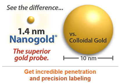
Nanogold® brings the versatility of fluorescent conjugation to gold labelling. Unlike colloidal gold preparations, Nanogold is small and highly uniform in size (1.4nm in diameter) to allow it to penetrate more antigens and provide better labelling. The Nanogold® particle is covalently and specifically linked to a hinge thiol on Fab’ or IgG. The conjugate therefore has excellent stability compared to colloidal gold-antibody preparations, reacting at specific sites under mild buffer conditions. Whilst the stoichiometry of conventional gold probes particles to IgG molecules varies from 0.2 to 10, Nanogold® preparations contain close to one Nanogold® to one Fab’ or IgG.
Key Features of Nanogold Conjugates
- The smallest commercially available gold immunoprobe.
- Penetrates and reaches antigens inaccessible to other probes: proven penetration up to 40 µm into cells and tissue sections.
- Extremely uniform 1.4 nm diameter gold label.
- Fab’, IgG and Streptavidin conjugates available.
- Close to 1 Nanogold® particle to 1 Fab’ (or IgG).
- Low non-specific affinity gives minimal background.
- Ultrasensitivity with silver enhancement: 0.1 pg of antigen detection on immunoblots.
- Gold is covalently attached to antibody remote from antigen binding region.
- High stability and long shelf life: conjugates show unchanged reactivity after storage for a year.
Stable to a wide range of pH and ionic strengths. - Label any molecule with a suitable reactive group: oligonucleotides, lipids, peptides, proteins, enzyme inhibitors and others. This is a big improvement over colloidal gold, which may be adsorbed only to antibodies and a limited range of proteins and peptides.
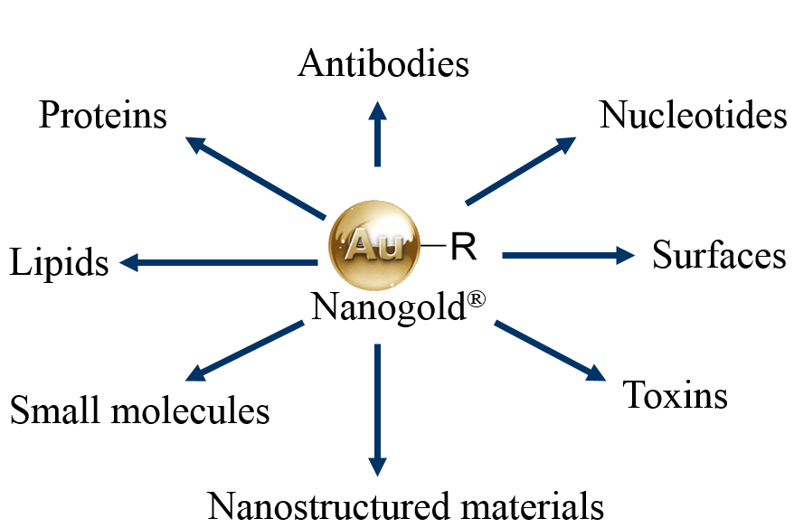
Applications for Nanogold Conjugates Include:
Gold labelling of protein fusion tags for electron microscopy
Electron microscopy of ultrathin cryosections
Pre-embedding electron microscopy
Post-embedding electron microscopy
Correlative electron and light/confocal microscopy
In situ hybridisation detection
Scanning electron microscopy
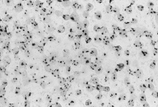
Fig 1. In Situ Hybridisation with Nanogold. Single copies of HPV 16 in SiHa cells detected by CARD-amplified Nanogold®-silver ISH. This cell line derived from cervical carcinoma contains only one to a few copies of HPV 16, which appear as single spots. Cytospin preparation, counterstained with Nuclear Fast Red (light micrograph courtesy of Dr. G. Hacker, Landeskrankenstalten, Salzbrg. Austria).
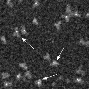
Fig 2. Electron Microscopy with Nanogold. This scanning transmission electron microscope (STEM) image clearly shows that labeling with Monomaleimido-Nanogold® (arrows) occurs specifically at a hinge thiol site on the IgG molecule.
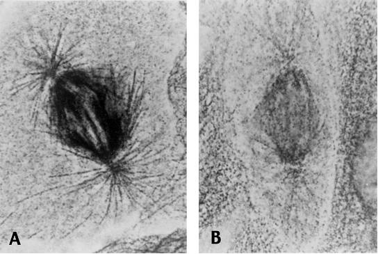
Fig 3. Light and Confocal Microscopy with Nanogold. Spindle microtubules labeled with anti-tubulin primary antibody followed by (LEFT) goat anti-mouse colloidal gold or (RIGHT) goat Fab’ anti-mouse-Nanogold (Light micrograph courtesy of Dr. D. Vandré and Dr. R. Burry, Ohio State University. Original magnification = 1300x).
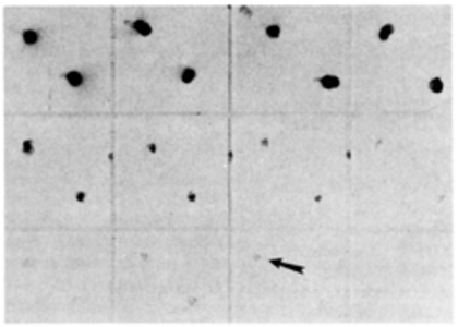
Fig 4. Immunoblot with Nanogold. Nanogold®-anti-mouse Fab’ blotted against mouse IgG, developed with LI Silver (Nanoprobes). These ultra small gold particles nucleate silver deposition so well that unprecedented sensitivity is achieved. This immunodot blot shows 0.1 pg sensitivity (arrow).
Nanogold products, manufactured by Nanoprobes are distributed in the UK & ROI by Caltag Medsystems. If you would like further information about their range, please contact us at techsupport@caltagmedsystems.co.uk or call us on 01280 827360.